New marker system leads to better implants

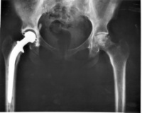
Hip-replacement surgery is a complex and exacting process. Currently, in order to ensure that patients receive the correct design and size of implant, orthopaedic surgeons perform a digital templating process that uses a single radiological scale marker to calibrate a radiograph.

The deployment of these single-scale markers, however, can be invasive and uncomfortable. Worse, the results obtained from them can be highly variable. That’s because a single marker – which often comprises a sphere mounted on a flexible arm – must be precisely positioned in the coronal plane of the hips and hence requires special expertise and time to apply correctly.

Now, a simple piece of equipment called KingMark, developed by Warwick University and University Hospitals Coventry and Warwickshire (UHCW) orthopaedic surgeon Richard King, in collaboration with Prof Damian Griffin at the university’s Warwick Medical School, is bringing a new level of accuracy and ease of use to the templating procedure.

The KingMark system consists of two separate radiographic markers instead of one. The first is a posterior marker consisting of a thin rectangular board with multiple radio-opaque metal marker rods arranged in series down its centre, while the second anterior marker comprises four radio-opaque steel balls contained within a flexible strap.

Prior to the radiography procedure, the rectangular board is placed underneath the patient’s pelvis, after which the strap containing the four anterior balls is placed in the midline over the patient’s suprapubic region. The radiograph is then taken.

To calculate the radiological magnification of the hips, the anterior marker ball and the posterior marker rod that lie most closely to an imaginary line connecting the hip centres are first identified on the radiograph.

The dimensions of the anterior and the posterior marker are then measured. This data can be used with the known size of both markers to calculate the magnification of the hips on the X-ray – with four times greater accuracy than the single-marker method.

’This enables us to routinely scale every pelvic X-ray that we perform with no difficulties for the radiographer or the patient. It also means that every time an X-ray is taken, the precise scaling information is provided on the radiograph,’ said King.

Damian Griffin, professor of trauma and orthopaedic surgery at Warwick Medical School, added: ’KingMark has removed the uncertainty about scaling digital radiographs for hip replacement. Our radiographers find it easy to use and I can be confident that the measurements I make on scaled radiographs are correct.’

To bring the new system to market, Warwick Ventures, which deals with the university’s intellectual property, approached Israel-based digital templating software specialist Voyant Health. The company immediately saw the potential for the product and agreed to manufacture and distribute the KingMark hardware.

The product has also been integrated with the company’s TraumaCad software, which can automatically detect the presence of the KingMark markers within an X-ray, calculate the magnification percentage and then size the X-ray image correctly.

The invention may also have potential for spinal work and trauma implants, as well as other joint-replacement operations.

The key facts to take away from this article

» KingMark utilises a posterior and an anterior radiographic marker
» It offers an accuracy four times greater than the single-marker method
» KingMark has removed the uncertainty of digital radiograph scaling
» It could also find use in other surgeries with magnification issues