This is the goal of a Glaxo-SmithKline-supported research programme at York University, where engineers have developed a way for transferring the magnetic spin of parahydrogen.
Through a reversible interaction with a specially designed molecular scaffold, the team has transferred parahydrogen’s magnetism to a range of molecules.
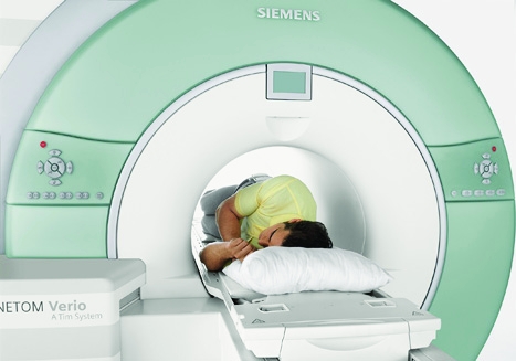
If this were applied to MRI, it could potentially increase sensitivity by giving scanners the ability to detect more molecules in the body than before.
Simon Duckett, a chemistry professor at York University, said that properly harnessed parahydrogen could help image biological processes, helping doctors spot tumours or signs of neurodegenerative diseases earlier.
Such diagnosis technology is possible with positron emission tomography (PET), where a patient is injected with a radioactive ’tracer’ material that mimics a biologically active molecule in the body, such as glucose. PET systems visualise whether the glucose was being metabolised by a tumour by detecting pairs of gamma rays emitted indirectly by the tracer.
MRI images are generated using radio waves to create changes in the magnetic field of hydrogen atoms in a sample. Often, these scans require magnetic contrast agents such as gadolinium. While not as harmful as PET tracer agents, they are heavy metal materials that can cause some health effects.
Duckett said that the new technique would, potentially, require that patients are only injected with contrast material endogenous to the body, such as amino acids.
’One of the benefits of the fact that the materials are endogenous to the body is you could repeat that measurement many times,’ he said. ’With PET, you wouldn’t want to be repeating the measurement many times because it is toxic.’ The technology also removes the need for large magnets and high magnetic fields.
This, added Duckett, coupled with new detector technology, means it is theoretically possible that these imaging systems could, one day, be shrunk down to a size that fits inside any GP office. It may even be possible to incorporate the detectors into a shirt that patients could wear for a scan.
’It relies on an awful lot coming together,’ he said. ’I would like to think we’re probably ready for clinical applications – depending on how quick things happen – in the next five to 10 years.’


Poll: Should the UK’s railways be renationalised?
I _do_ remember British Rail - and that it was <i>literally</i> a national joke https://youtu.be/zV2lmSDKvO8