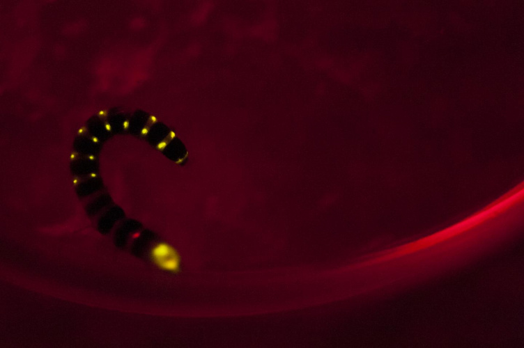
Many animals have the ability to emit light, but the most studied emit yellow-green light. An exception is the railroad worm, the larva of the Phrixothrix hirtus beetle, which emits both red and yellow-green light. It has a row of light-emitting organs along its body, giving rise to an effect like the lights of an illuminated train carriage, hence the name. An article in Scientific Reports details research by engineers and biologists at the Federal University of Sao Carlos and the University of electro-Communications in Tokyo to determine the mechanism of the red light emission.
The Brazilian team, attached to the country’s National Energy and Materials Research Centre, built on previous research by principal investigator Vadim Viviani which showed that luciferase, an enzyme involved in the production of light by invertebrates, when taken from fireflies changed the colour of the light they emitted from green to red in a test tube in response to the acidity of the medium in which the light-emitting reaction took place. However, they still did not know how the railroad worm naturally emitted red light. This effect is confined to a cell on the larva’s head, producing a light which the larva uses to find its way in the dark (the row of yellow lights along its side frighten away predators).
_________________________________________________________
Further reading
- Inhalable therapy may produce drugs in the lungs
- Researchers develop firefly-inspired glowing nanorods
- Real-time cell activity
_________________________________________________________
Researchers in this area has developed methods to clone different luciferases, and the Tokyo team synthesised red light-emitting analogues of a related protein, luciferin, which Viviani’s colleague Vanessa Rezende Bevilaqua tested using luciferases cloned by the Brazilian group from both fireflies and railroad worms. Bevilaqua found that larger luciferins interacted better with the railroad worm luciferase, emitting red light more efficiently. "The luciferases that catalyze green and yellow light have a small cavity and therefore don't bind well to the large-structure luciferin analogs, which have very little luminescent activity," Viviani said. "On the other hand, these large analogs interact well with luciferases that catalyze red light. We deduced from this that railroad worm luciferase has a large active site cavity capable of binding to the analogues."
This could be useful in medical imaging because in mammals, blood and muscular cells do not absorb red light, so luciferase-based techniques increasingly used to visualise processes taking place involving these cells do not work. This means that researchers cannot easily study processes taking place in bone or haemoglobin-rich tissues. "When these substances are examined with conventional luciferase that emits green, yellow or blue light, it's impossible to see biochemical and pathological processes clearly because pigments such as haemoglobin and myoglobin absorb most of the light in these parts of the chromatic spectrum," Viviani said. Imaging using railroad worm luciferase instead would throw light on these processes, assisting in the study of diseases and screening of drugs.


Nanogenerator consumes CO2 to generate electricity
Nice to see my my views being backed up by no less a figure than Sabine Hossenfelder https://youtu.be/QoJzs4fA4fo