US scientists have created an imaging device with a layout that is based on the human eye.
The researchers, at the University of Illinois and Northwestern University, developed the hemispherical 'eye' camera using an array of single-crystalline silicon detectors and electronics, configured in a stretchable, interconnected mesh.
John Rogers, the Flory-Founder chair professor of Materials Science and Engineering at the University of Illinois, said: 'Conformally wrapping surfaces with stretchable sheets of optoelectronics provides a practical route for integrating well-developed planar device technologies onto complex curvilinear objects.
'This approach allows us to put electronics in places where we couldn’t before. We can now, for the first time, move device design beyond the flatland constraints of conventional wafer-based systems.'
The camera’s design is based on that of the human eye, which has a simple, single-element lens and a hemispherical detector. The camera integrates such a detector with a hemispherical cap and imaging lens, to yield a system with the overall size, shape and layout of the human eye.
To make the camera, the researchers begin by moulding a thin rubber membrane in the shape of a hemisphere. The rubber membrane is then stretched to form a flat drumhead.
Next, a prefabricated focal plane array and associated electronics - created by conventional planar processing - are transferred from a silicon wafer to the tensioned drumhead membrane.
When the tension is released, the membrane returns to its original shape. This process compresses the focal plane array, causing specially designed electrical interconnects to delaminate from the rubber surface and form arcs, pinned on the ends by detector pixels.
These deformations accommodate strains associated with the planar to hemispherical transformation, without stressing the silicon, as confirmed by mechanics modeling performed by researchers at Northwestern.
The array package is then transfer printed to a matching hemispherical glass substrate. Attaching a lens and connecting the camera to external electronics completes the assembly.
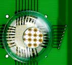
Schematic illustration of the steps for using compressible silicon focal plane arrays and hemispherical, elastomeric transfer elements to fabricate electronic eye cameras
Over the last 20 years, many research groups have pursued electronic eye systems of this general type, but none has achieved a working camera.
Rogers, who is also a researcher at the Beckman Institute and at the university’s Frederick Seitz Materials Research Laboratory, added: 'Optics simulations and imaging studies show that these systems provide a much broader field of view, improved illumination uniformity and fewer aberrations than flat cameras with similar imaging lenses.
'Hemispherical detector arrays are also much better suited for use as retinal implants than flat detectors. The ability to wrap high-quality silicon devices onto complex surfaces and biological tissues adds very interesting and powerful capabilities to electronic and optoelectronic device design, with many new application possibilities.'


IEA report claims batteries are ‘changing the game’
Batteries come with major weight penalties in vehicles and to date have only achieved very limited application in the rail sector where range and...