
Microvesicles shed by cancer cells are even more numerous than those released by normal cells, so detecting them could prove a simple means for diagnosing cancer.
In a study published in Nature Medicine, investigators at the Massachusetts General Hospital (MGH) Center for Systems Biology (CSB) demonstrate that microvesicles shed by brain cancer cells can be reliably detected in human blood through a combination of nanotechnology and their new NMR-based device.
‘About 30 or 40 years ago, people noticed something in the bloodstream that they initially thought was some kind of debris or “cell dust”,’ said Hakho Lee of the CSB and co-senior author of the study with Ralph Weissleder, director of the CSB. ‘But it has recently become apparent that these vesicles shed by cells actually harbour the same biomarkers as their parent cells.’
Circulating tumour cells (CTCs) have been regarded as a potential key to improved cancer diagnosis, but Lee explained in a statement that CTCs are extremely rare and finding them in blood is difficult.
Microvesicles, however, are abundant in the circulation and, unlike CTCs, are small enough to cross the blood/brain barrier, which means that they could be used to detect and monitor brain cancers, he added.
Glioblastoma multiforme (GBM) is the most common and most aggressive brain cancer in humans and by the time it is diagnosed patients typically have less than 15 months to live.
One of the biggest challenges with this condition is accurate disease monitoring to establish whether patients are responding to treatment.
Currently, the only way to diagnose and monitor GBM is with biopsies and imaging tests, making long-term treatment monitoring difficult, invasive and impractical.
To address this need, the CSB team sought to develop a simple blood test that could be used to easily monitor disease progression.
‘The issue with microvesicles, however, is that they are very small, so there are not many technologies out there that can detect and molecularly profile them,’ said Lee. ‘That is where our new technology comes in.’
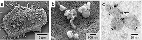
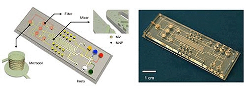
By using nanotechnology to magnetically label microvesicles, and by adapting and improving equipment they developed last year to detect cancer cells with a miniature, handheld NMR device, the MGH researchers were able to detect the tumour microvesicles in blood samples from mice bearing human GBM tumours and eventually in samples from human GBM patients.
Compared with other gold-standard techniques, this new technology is said to have demonstrated excellent detection accuracy.
However, unlike other methods — which can be time consuming and require much greater sample volumes as well as expertise to perform — NMR detection is quick and simple, potentially providing almost instant results from a small blood sample in a doctor’s office.
The MGH CSB team is currently extending this platform to other types of cancer and to other diseases such as bacterial infection. A number of clinical studies are currently ongoing and others are in the planning stages, with the goal of eventually commercialising the technology.
Poll: Should the UK’s railways be renationalised?
Rail passenger numbers declined from 1.27 million in 1946 to 735,000 in 1994 a fall of 42% over 49 years. In 2019 the last pre-Covid year the number...