Watching cells is tricky. To determine how they behave in a living body, they must be viewed in an environment as close to natural as possible; and that means with the other cells that live alongside them. Light interacts differently with different shapes and substances, so conventional microscopy can only produce blurred and confusing images. Isolating the cells on a glass slide will not give information about their behaviour, and specialised microscopy using powerful forms of light can damage their delicate structure.
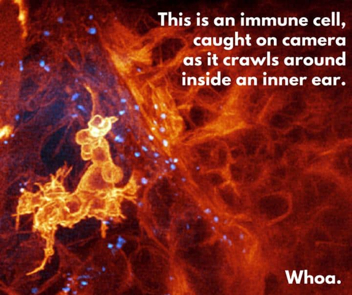
Image (and text): Boston Children's Hospital
Researchers at the Howard Hughes Medical Institute in Virginia, working with Boston Children’s Hospital and Harvard Medical School, have devised a combination of technologies that produces startlingly sharp results.
The team, led by Dr Eric Betzig, co-winner of the 2014 Nobel Prize for chemistry for his role in developing super-resolved fluorescence microscopy, used a form of microscopy known as lattice light-sheet, which illuminates the subject by scanning a thin sheet of light repeatedly across living tissue at high speed to observe cells inside an embryonic zebrafish, suitable for this type of work because it has translucent skin.
To avoid the problem of distortion of the image, Betzig borrowed a technique from large, ground-based telescopes: adaptive optics. In telescopes, this is used to overcome the distorting effects of the Earth’s atmosphere. A laser is used to project a “guide star” into the telescope’s field of view, and its distortion is analysed and used to change the shape of one of the mirrors in the telescope’s optics to produce a sharp image. The microscopy team used a similar technique, passing a laser through the target tissue and comparing its appearance on either side. As with the telescope, this allows the microscope to correct distortions in the image.
Describing its research in Science, the team explains how it used the technique to look at immune cells inside the inner ear of the zebrafish. “Every time we've done an experiment with this microscope, we've observed something novel and generated new ideas and hypotheses to test," said Tom Kirchhausen, a co-author on the paper who works at the Children’s Hospital and at Harvard. "It can be used to study almost any problem in a biological system or organism I can think of." Zebrafish can be genetically modified to study human disease models, the team has also used the technique to watch metastatic human breast cancer cells move around inside the fish’s bodies.
The team is now working on miniaturising the equipment needed to use the technique, which currently occupies a 3m long table. Betzig believes that adaptive optics has great potential in microscopy, and without it subcellular imaging is “just too damn fuzzy.”
See a crawling immune cell here:


Poll: Should the UK’s railways be renationalised?
I think that a network inclusive of the vehicles on it would make sense. However it remains to be seen if there is any plan for it to be for the...