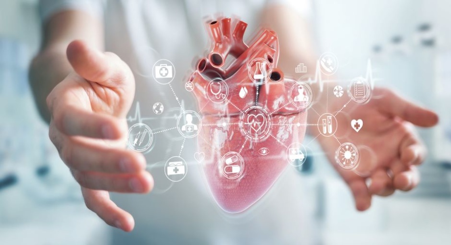
The technology, developed by researchers from the School of Biomedical Engineering & Imaging Sciences at Kings College London and the Evelina London Children’s Hospital, brings together scans that are routinely used to plan congenital heart disease surgery to create a three-dimensional, beating digital twin of the heart.
MORE FROM MEDICAL & HEALTHCARE
The researchers hope that using VR to plan and practice procedures will shorten operating times and reduce the need for multiple surgeries, leading to better outcomes and experiences for patients and their families. They hope that it could be in regular use within the next two years.
“We have had a lot of help from the fantastic team at King’s Medical Engineering Quality Management System who are helping us to move the device through from a prototype to a nationally regulated device which can be used to help plan these complex procedures,” said Dr Natasha Stephenson, Clinical Research Fellow in Congenital Cardiovascular Imaging at the School of Biomedical Engineering & Imaging Sciences.
Trials of an early version of the technology, which used only ultrasound scans of the heart to create the VR heart, found that surgeons preferred it for understanding the anatomy of their patient’s hearts. They also reported that it increased their confidence and improved their decision making.
Funding from the British Heart Foundation and Evelina London Children’s Charity has supported the researchers in introducing computed tomography (CT) and magnetic resonance imaging (MRI) scans into the system. While these types of scans are regularly used to help plan surgeries, they are usually only viewed on a flat screen.
Now, surgeons using the technology are immersed into the heart, allowing them to interact with and manipulate the images. They can also test options for the procedure in VR before they get to the operating table.
“Procedures to repair the heart’s anatomy can be complex, and surgeons don’t like surprises,” said lead researcher Professor John Simpson, Professor of Paediatric and Fetal Cardiology at Evelina London and King’s College London. “Our technology will allow surgeons to plan and practice these procedures, and we’re currently applying for approval for it to be used in this way.”
He continued: “We think that this technology could also be used outside of congenital heart disease surgeries, to plan any procedure which aims to correct a structural problem within the heart, such as valve surgery in an adult patient.”


Virgin Atlantic’s Flight100 saved 95 tonnes of CO2 in first SAF flight
Good comment. I think these reports are different from many others , in that they were prepared outside the government and the issues, they raised, of...