UK team uses AR to assist reconstructive surgery
An Imperial College London team working at St Mary's Hospital has used augmented reality (AR) to pinpoint blood vessels during reconstructive surgery.
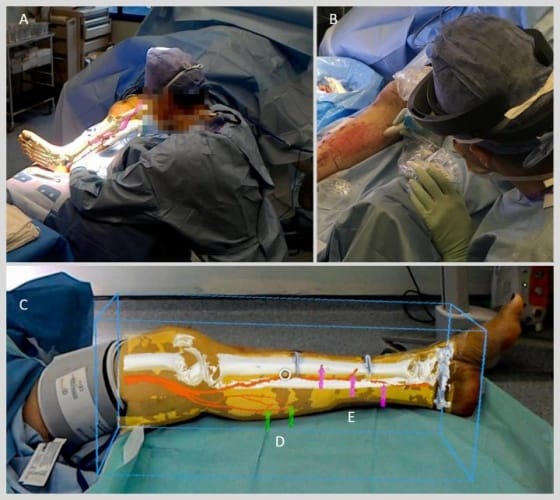
(Credit: 'Philip Pratt, et al. Eur Radiol Exp, 2018)
In what is claimed to be a world first, images of CT scans showing patients’ bones and blood vessels were overlaid on lower limbs using the Hololens, a ‘mixed reality’ visor developed by Microsoft. The technique assisted surgeons in attaching fasciocutaneous flaps – grafts containing skin and blood vessels – to patients that had suffered major trauma, ensuring oxygenated blood was flowing to the grafted skin.
"We are one of the first groups in the world to use the HoloLens successfully in the operating theatre," said Dr Philip Pratt, a research fellow in the Department of Surgery & Cancer and lead author of the study, published in European Radiology Experimental.
"Through this initial series of patient cases we have shown that the technology is practical, and that it can provide a benefit to the surgical team. With the Hololens, you look at the leg and essentially see inside of it. You see the bones, the course of the blood vessels, and can identify exactly where the targets are located."
Register now to continue reading
Thanks for visiting The Engineer. You’ve now reached your monthly limit of news stories. Register for free to unlock unlimited access to all of our news coverage, as well as premium content including opinion, in-depth features and special reports.
Benefits of registering
-
In-depth insights and coverage of key emerging trends
-
Unrestricted access to special reports throughout the year
-
Daily technology news delivered straight to your inbox


INWED Engineering Profile: Naval Architect Ellie Driver
Not a woman I´d want to cross … oh, that was Elle Driver