This is the aim of a research programme at the Wellcome Laboratories for Vision Sciences at City University London.
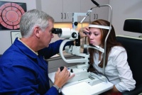
A team led by Steve Gruppetta, an academic fellow in the Department of Optometry and Visual Science, is developing a high-resolution 3D retinal imaging instrument that is inexpensive and compact enough for use in any optometrist or GP office. The machines usually used by optometrists and GPs are inexpensive but produce low-resolution 2D images.
Gruppetta and his team believe the new instrument will give the medical community a better opportunity to spot structural changes to a patient’s retina. This could help lead to the earlier diagnosis of diseases such as glaucoma, which could then be treated before a patient suffers impaired vision.
Gruppetta is waiting for patent approval before divulging full details on how the device works. He did reveal, however, that it would be based on a form of microscopy.
According to Gruppetta, retinal imaging instruments based on microscopy already exist, but they are bulky, expensive and usually found in hospitals. These instruments, which include scanning laser ophthalmoscope (SLO) and optical coherence tomography (OCT), are made with complex moving mirrors and expensive light sources, including superluminescent diodes or femtosecond lasers.
SLO and OCT build up 3D images by scanning laser light horizontally and vertically on a section of a patient’s retina. Gruppetta said this is accomplished using oscillating mirrors – one horizontally and the other vertically – which send light across the retina.
In the case of SLO, the light goes through an optical system comparable to those used in confocal microscopy. In this case, a magnified image is acquired by using a confocal ’pinhole’ to eliminate out-of-focus light. An OCT system, on the other hand, uses an interferometer and the imaging is based on the interference of light. Both of them include a detector for collecting light, which is digitally reconstructed point by point into a 3D representation of the retina.
The key challenges for the team will be developing an instrument with cheaper light sources and fewer moving parts. Another challenge will be reducing the complexity of the computational processing required to form a digital 3D image.
Gruppetta envisions such systems being available within several years following clinical trials.


Water Sector Talent Exodus Could Cripple The Sector
Well let´s do a little experiment. My last (10.4.25) half-yearly water/waste water bill from Severn Trent was £98.29. How much does not-for-profit Dŵr...