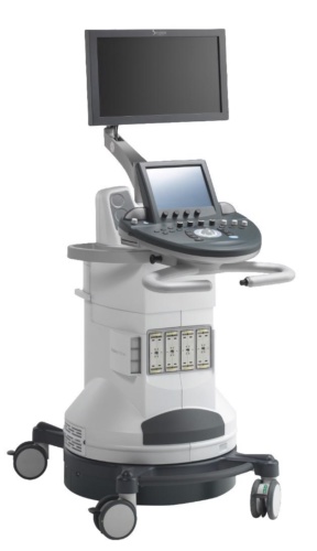
New breast imaging tech can tell malignant from benign
European scientists have developed a new breast imaging technique that can distinguish malignant from benign tumours, potentially ending unnecessary biopsies.

The Horizon2020 project SOLUS combines imaging and ultrasound techniques, with patients examined using a non-invasive pen probe, similar to that used for pregnancy scans. Using a technique called ‘diffuse optical imaging’, the probe can monitor changes in concentrations of oxygenated and deoxygenated haemoglobin, collagen, lipids and water present in a suspected tumour against a pre-programmed set of results. It’s these parameters that indicate whether a tumour is cancerous or not.
“We have been applying diffuse optics to different aspects of breast cancer (lesion discrimination and an estimate of cancer risk) for 20 years now,” said Professor Paola Taroni from Politecnico di Milano, Italy.
Tumour imaging technique promises cancer care boost
Infrared imaging shines light on biomarkers in cancer cells
“Our team developed a multi-wavelength time-domain optical mammography which was used in clinical studies on more than 400 patients. Now an upgraded version has just entered clinics for the monitoring of neoadjuvant chemotherapy of breast cancer. Furthermore, our group performed a lot of work on other diagnostic applications, such as functional brain imaging and muscle oximetry.”
Register now to continue reading
Thanks for visiting The Engineer. You’ve now reached your monthly limit of news stories. Register for free to unlock unlimited access to all of our news coverage, as well as premium content including opinion, in-depth features and special reports.
Benefits of registering
-
In-depth insights and coverage of key emerging trends
-
Unrestricted access to special reports throughout the year
-
Daily technology news delivered straight to your inbox


Water Sector Talent Exodus Could Cripple The Sector
Maybe if things are essential for the running of a country and we want to pay a fair price we should be running these utilities on a not for profit...