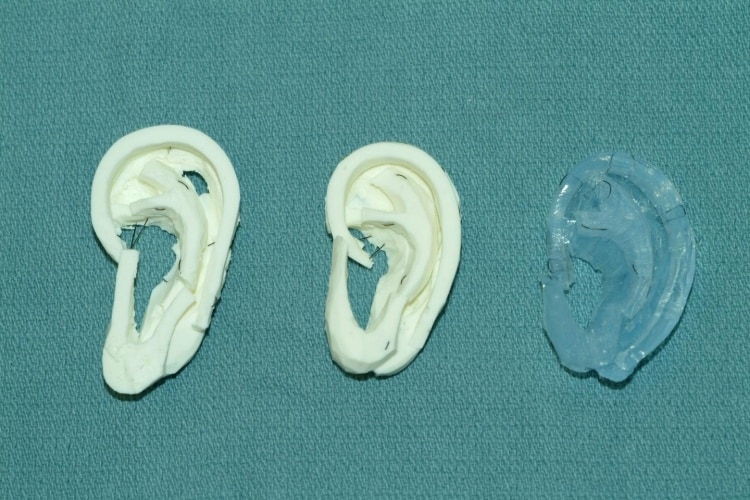
When creating a new ear for patients with missing or underdeveloped ears, surgeons use cartilage taken from the rib, carving an ear shape from it. Typically, a variety of different materials are used to practise the technique, including soap, vegetables and dental moulds. Using a homemade material, the UW team was able to 3D-print a more lifelike model of the rib cartilage for surgeons and trainee surgeons to practise on.
“It’s a huge advantage over what we’re using today,” said Angelique Berens, otolaryngology resident at UW and lead author of the research paper.
“You literally take a bar of Lever 2000 while the attending is operating and you carve ear cartilage. It does teach you how to get the shape right, but the properties are not super accurate – you can’t bend it, and sewing it is not very lifelike.”
The results of the study are published in an abstract that was presented this week at the American Academy of Otolaryngology – Head and Neck Surgery conference in Dallas, Texas. Three experienced surgeons tested the 3D-printed cartilage against more expensive dental impression material, comparing their firmness, feel and suturing quality to real rib cartilage. All agreed that the UW material was a better proxy for real cartilage, recommending that it be introduced as a training tool.
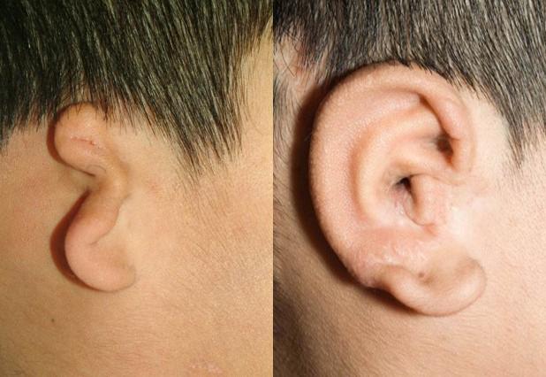
Another advantage is that the UW models are printed from a CT scan, and can mimic an individual’s unique anatomy. This means that difficult surgery with unorthodox elements can be practised ahead of time. The material itself was arrived at following a process of trial and error with combinations of silicone, corn starch, mineral oil and glycerine to replicate human tissue that the lab’s surgical robot could manipulate.
“I would go to the craft store and Home Depot and say I want to make models – what aisle should I go to?” said co-author Sharon Newman, who recently graduated from UW with a bioengineering degree.
“It turns out a lot of these ideas were based off of materials people use for arts and crafts like rings or other jewellery.”
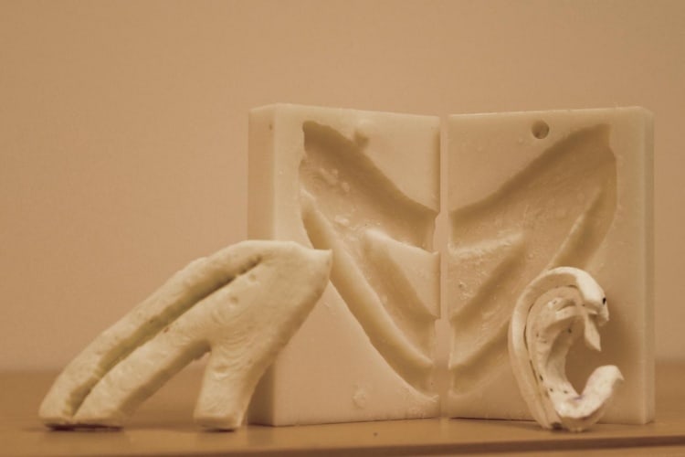
As everyday materials are used to create the models, large numbers of units can be fashioned from one mould, meaning the surgery could be much more widely practised.
“With one 3-D printed mold, you can make a billion of these models for next to nothing,” said Berens. “What this research shows is that we can move forward with one of these models and start using it.”


Poll: Should the UK’s railways be renationalised?
Rail passenger numbers declined from 1.27 million in 1946 to 735,000 in 1994 a fall of 42% over 49 years. In 2019 the last pre-Covid year the number...