X-ray imaging technique could unlock small crystal properties
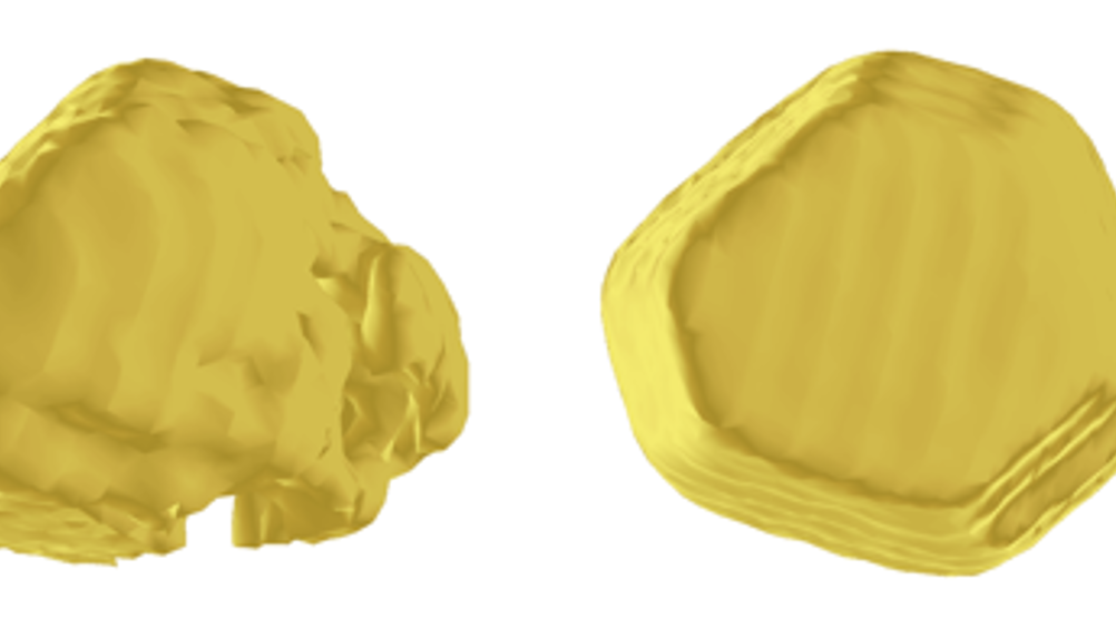
A new X-ray imaging technique could help materials scientists unlock the properties of the smallest crystals, enabling faster electronic devices and possibly even stronger cement.

Using computing techniques that model the radiation from the X-ray source, a team from the London Centre for Nanotechnology at UCL revealed the shape of gold nanocrystals in more detail than ever before.
‘Gold is really just a model material for these studies; we’ve been looking at crystals of metal oxides, steels and cement,’ said Prof Ian Robinson, who has written a paper on the research for Nature Communications with Jesse Clark. ‘Looking at the smallest sizes of crystals is a good way to tackle questions about how cement works, for example, and where these materials’ properties come from.’
For example, he said, the strength of metals depends on stopping the materials’ crystal grains from sliding against each other. ‘One of the things that makes nanomaterial such an exciting prospect is that smaller is stronger,’ Robinson said. ‘Our imaging method will allow us to see the strain in the grains as they interact with each other, which will help us to find ways to use that strength.’
Register now to continue reading
Thanks for visiting The Engineer. You’ve now reached your monthly limit of news stories. Register for free to unlock unlimited access to all of our news coverage, as well as premium content including opinion, in-depth features and special reports.
Benefits of registering
-
In-depth insights and coverage of key emerging trends
-
Unrestricted access to special reports throughout the year
-
Daily technology news delivered straight to your inbox


Water Sector Talent Exodus Could Cripple The Sector
One possible reform to the Asset Management Plan (AMP) system would be to stagger the five year cycle across the ten or so water businesses, so that...