Researchers at the University of Southern California's Viterbi School of Engineering have successfully demonstrated a novel High-resolution Ultrasonic Transmission Tomography (HUTT) system that offers 3D images of soft tissue that are superior to those produced by existing commercial X-ray, ultrasound or MRI units.
Vasilis Marmarelis, a professor of biomedical engineering at the Viterbi School, presented HUTT images of animal organ tissue in San Diego at the 28th International Acoustical Imaging Symposium on March 21st.
According to Marmarelis, HUTT offers nearly order-of-magnitude improvement in resolution of structures in soft tissue (i.e., 0.4 mm, compared to 2 mm for the best alternatives).
Several other features promise to make the technology a scientific and clinical tool of great power.
Algorithms developed by his team enable HUTT to differentiate separate types of tissue based on their distinctive “frequency-dependent attenuation” profiles that should allow clinicians to distinguish malignant lesions from benign growths in a non-invasive and highly reliable manner.
Scans can be performed in a matter of a few minutes and because they are ultrasonic, they do not use potentially harmful ionizing radiation.
The system requires a minimum of special pre-scan procedures and appears likely, in clinical use, to be more comfortable for patients than alternatives.

HUTT image of kidney tissue
In traditional hand-held ultrasound systems, sound waves are broadcast into the tissue, and the echoes produce an image of the reflecting interfaces. The sound transmitter and the receiver in such systems are both on the same side of the sample. However, only a tiny fraction of the transmitted sound comes back as echo on soft tissues, while a much larger fraction (about 2000 times bigger) is transmitted through the soft tissue.
The HUTT system, however, collects the sound transmitted through the tissue, allowing the formation of better images with greater clarity and resolution. To do so, it transmits an extremely short ultrasonic pulse (about 250 nanosecond) of 4-12 megahertz frequency (far above human hearing) and picks up the pulse on the other side after it has travelled through the imaged object.
The transmitted pulses come from an array of very small ultrasonic transducers of sub-millimetre dimensions. A parallel array of transducers on the other side receives the pulses after they travel through the imaged tissue.
The two arrays, transmitter and receiver, are mounted on opposite sides of a drum that spins as it rises around the object (which is suspended in water), creating a stack of tomographic image slices which the software algorithms turn into 3D images. A sophisticated coding/decoding signal scheme then allows the system to distinguish which signal came from which transducer.
When the transducer captures the signal, it is processed with advanced signal processing algorithms, specially developed by Marmarelis’ group, to form multi-band images.
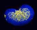
Choosing a target. Different tissues in the same organ (here, a sheep kidney) vary in "attenuation" characteristics: how much ultrasound of various frequences they let through. Algorithms that incorporate empical findiings of these differences can quickly discriminate between tissue types in scans, and even digitally apply "dyes" to the captured image to make a given type of tissue stand out. From left to right, 1: overall view. 2. capsule (blue) 3. blood vessels (red) 4. papillary ducts, (magenta) 5. calyces (green).
The most critical feature of the HUTT imaging technology is its potential to reliably differentiate types of tissue based on their multi-band signatures caused by their varying attenuation patterns. This promises to allow non-invasive detection of lesions in clinical diagnosis, which represents the “holy grail” of medical imaging.
The team found it possible to identify various anatomical structures within the kidney based on their distinctive attenuation characteristics, so that computerized algorithms could display in colour-coded fashion one tissue in red, another in green, and so forth.
The technology could also be used to isolate one type of tissue, allowing, for example, all the blood vessel structures to be displayed alone and studied. In addition to improved resolution, the system can locate tissue features with extreme precision in a objective, fixed-coordinate 3D grid, crucial for guiding surgical procedures.


Poll: Should the UK’s railways be renationalised?
All public service companies should be nationalised for many different reasons, particularly railways, not the least because the tax payer has already...